Do you need an MRI

Why are MRIs Futile in most Back Pain Cases?
MRIs provide detailed pictures of the spine, allowing visualization of bones, discs, nerves, and other tissues. While this detailed view can be invaluable in diagnosing certain conditions, it can also be misleading in back pain cases. MRIs frequently fail to provide meaningful insights or improve patient outcomes in cases of back pain.
Let us first understand what MRIs are for
MRIs are more advanced as imaging techniques compared to X-rays, providing detailed views without using radiation, unlike CT scans. Additionally, MRIs are non-invasive and widely accessible. They provide detailed images of soft tissues, including the spinal cord, intervertebral discs, and surrounding structures. Physicians often prescribe MRIs to pinpoint the source of back pain, assuming that visualizing anatomical abnormalities will lead to effective treatment strategies. It has come a long way from hazy images of Tesla 1 to Tesla 3.5 but it may not reveal the exact reason for pain in most of the cases.
Limitations in MRI Report Specific to Lower-Back Pain Diagnosis
- MRIs are performed in the supine position (lying on the back). In most of cases, the pain is not nonexistent while lying down, it only starts in sitting or standing positions as these postures put more weight on the discs. Hence, MRIs do not capture the discs under stress.
- They may not be the best tool for diagnosing muscle stiffness or shortening. Imaging quality differs with various updates of MRIs Tesla 1, Tesla 1.5, and Tesla 3 3.5.
- MRIs cannot determine the age of a herniated disc.
- It mostly gives a similar image even if done one year apart. Although patients experience some painful days and some pain-free days, the MRI images are pretty much the same.
MRI: A Double-Edged Sword
- High Rate of False Positives: Studies have shown a surprisingly high percentage of asymptomatic individuals (people without back pain) having abnormal MRI findings [1].
- Anxiety and Over-treatment: An abnormal MRI result can be a source of anxiety for patients, leading them to believe their condition is severe and requires surgery, and hindering recovery by focusing on the visualized abnormality rather than addressing the pain itself [2].
- Unnecessary Costs: MRIs are expensive, and the cost often outweighs the benefit in uncomplicated back pain [3].
The Pitfalls of Overdiagnosis
The identification of structural abnormalities can be inconsequential to the patient's symptoms. When MRI reveals disc herniations, degenerative changes, or spinal stenosis in individuals experiencing back pain, clinicians may attribute the symptoms to these findings. However, growing evidence suggests that these structural anomalies often exist independently of pain or are unrelated to the patient's symptoms. Consequently, overreliance on MRI findings may lead to unnecessary interventions, including surgery, with limited benefit or even exacerbation of symptoms.
The Complexity of Pain
Pain is a multifaceted phenomenon influenced by biological, psychological, and social factors. While structural abnormalities may contribute to pain in some cases, they often do not fully account for the complexity of back pain. Factors such as psychosocial stressors, maladaptive pain processing, and poor coping mechanisms can significantly influence the experience and persistence of symptoms. MRI, despite its prowess in anatomical visualization, cannot capture these nuanced aspects of pain.
The Need for a Paradigm Shift
The limitations of MRI in back pain cases underscore the necessity of a paradigm shift in clinical practice. Rather than relying solely on imaging findings, a comprehensive approach to back pain management should incorporate patient history, physical examination, psychosocial assessment, and shared decision-making.
The Takeaway: A Collaborative Approach
The decision to get an MRI should be a collaborative one between you and your doctor. By understanding the limitations of MRIs and exploring conservative treatment options first, you can avoid unnecessary costs, anxiety, and potentially invasive procedures. Back pain is often a complex issue, and the best approach is a multi-pronged one that addresses the underlying cause and promotes long-term healing. Discuss your specific situation with your doctor to create a personalized treatment plan that prioritizes both effectiveness and cost-effectiveness for managing your back pain.
References:
1. Active Health KC. (n.d.). Have low back pain or neck pain? An MRI might not be a good idea [Active Health KC]. Retrieved April 18, 2024, from [activehealthkc.com]
2. Same reference as #1
3. Chou, R., Qaseem, A., Owens, D. K., Loeser, W. F., & Cifu, A. S. (2018). Diagnosis and Treatment of Low Back Pain: A Joint Clinical Practice Guideline from the American College of Physicians and the American Pain Society. Annals of Internal Medicine, 169(7), 477–483. Wood R, Bassett K, Foerster 5th, Spry C, Tong L. 1.5 tesla magnetic resonance imaging scanners compared with 3.0-tesla magnetic resonance imaging scanners: a systematic review of clinical effectiveness. CADTH Technol Overv. 2012;2(2):e2201. Epub 2012 Jun 1. PMID: 23002376; PMCID: PMC3442613.
4. Brinjikji, W., Luetmer, P. H., Comstock, B., Bresnahan, B. W., Chen, L. E., Deyo, R. A., ... & Jarvik, J. G. (2015). Systematic literature review of imaging features of spinal degeneration in asymptomatic populations. American Journal of Neuroradiology, 36(4), 811-816.
5. Chou, R., Fu, R., Carrino, J. A., Deyo, R. A., Clinical Guidelines Committee of the American College of Physicians. (2009). Imaging strategies for low-back pain: systematic review and meta-analysis. The Lancet, 373(9662), 463-472.
6. Jenkins, H. J., & Hancock, M. J. (2019). Imaging for low back pain: is clinical use consistent with guidelines? A systematic review and meta-analysis. Spine Journal, 19(7), 1256-1272.
7. Maher, C., Underwood, M., & Buchbinder, R. (2017). Non-specific low back pain. The Lancet, 389(10070), 736-747.
.png)
Editorial issue 9 -
Long ago, a young disciple asked his Guru, "Master, if yoga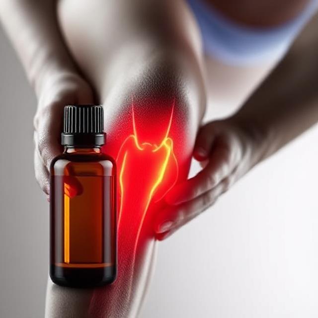
Natural Relief | Essential Oils for Knee Pain & Inflammation Management
The knee joint is one of the strongest joints of a human bod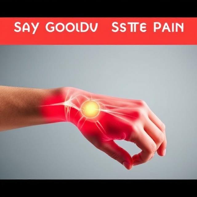
Say Goodbye to Wrist Pain: How to Prevent Carpal Tunnel and Mouse Arm
We all know the feeling — after hours of typing or us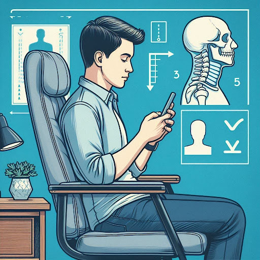
How Does Ergonomics impact Joint health such as neck and spine?
Ergonomics refers to how your body responds to daily activit